
If you are over 40, you might notice your morning walk feels a bit stiffer than it used to. Your stride feels shorter, your knees ache after a brisk stroll, or your balance feels subtly off. This guide will teach you how to unlock 5 ankle mobility steps if you’re 40+.
Learn how to stay fit after 40: 6 Strong Hybrid Movements for 40+ Adults
Many active adults blame these issues on knee joint degeneration or general aging, but the true culprit often lives lower down in the ankle complex.
Ankle dorsiflexion—the ability of your shin to travel forward over your foot—is the foundation of efficient upright movement. When ankle mobility drops, your body adapts by altering your stride, forcing your lower back, hips, and knees to absorb unnatural forces.
Unlocking ankle mobility is not about performing endless generic stretching routines. It requires systematic assessment, clear differentiation of movement restrictions, and progressive movement training. Here are five steps to restore your ankle mobility and reclaim your stride power.
The Science of Over-40 Ankle Mechanics
Biomechanical research shows that passive ankle range of motion naturally decreases with age, particularly in the dorsiflexion pathway.
When ankle dorsiflexion is restricted during gait:
-
Forward Propulsion Drops: The tibia cannot roll smoothly forward over the foot, reducing hip extension power during push-off.
-
Joint Loading Increases: The knee compensates by rotating inward or over-extending, which transfers excess stress up the kinetic chain.
-
Balance & Stability Decline: Decreased range of motion impairs postural stability, increasing the energy cost of walking and raising fall risks.
Restoring functional ankle mobility directly supports joint health, gait efficiency, and long-term movement confidence.
STEP 1: Assess Your Baseline Ankle Dorsiflexion
Before attempting any corrective drills, you must objectively test your current range of motion to establish a baseline.
Many individuals falsely assume their ankles are restricted when the actual issue stems from movement apprehension or poor coordination.
Half-Kneeling Wall Screen
-
Setup: Stand barefoot facing a wall in a half-kneeling position, placing your front foot perpendicular to the wall.
-
Measurement: Position your fist between your big toe and the wall (approximately 3.5 to 4 inches away).
-
Execution: Keep your front heel glued flat to the floor. Drive your front knee straight forward, trying to touch the wall.
-
Checkpoint: If your knee touches the wall without your heel lifting, you possess functional dorsiflexion. If your heel lifts or your knee cannot reach the wall, you have identified a restricted baseline.
STEP 2: Differentiate Soft Tissue vs. Joint Restriction
Ankle restrictions fall into two distinct biomechanical categories: soft tissue tightness or joint capsules/articular restrictions. Treating a joint restriction with soft tissue foam rolling will yield little progress.
The End-Range Feel Test
-
Setup: Stand barefoot on an elevated step or curb with the ball of one foot firmly on the edge.
-
Straight-Knee Test: Lower your heel toward the floor with a straight knee. Hold for 5–10 seconds. Note where you feel the sensation: a stretch in the calf/Achilles region, or a pinching block at the front of the ankle.
-
Bent-Knee Test: Bend your testing knee slightly and lower your heel again. Note the sensation.
-
Analysis: A stretch in the back of the leg across both positions indicates a soft tissue restriction (calves or soleus). A hard blocking or pinching sensation at the front of the ankle indicates a joint restriction.
STEP 3: Address Soft Tissue Restrictions
If your assessment revealed calf or Achilles tightness, address the tissue quality and follow up with structured, controlled stretching.
Targeted Tissue Release & Bi-Phasic Stretching
-
Deep Calf Release: Use a soft lacrosse ball or dense foam roller under the belly of the calf (gastrocnemius and soleus). Find a sensitive spot and pause, slowly flexing your foot up and down for 45–60 seconds.
-
Straight-Knee Bi-Phasic Stretch: Stand facing a wall, extend one leg back with a straight knee, and press the heel down. Perform small, controlled oscillations forward for 30 seconds, then hold statically for 60 seconds.
-
Bent-Knee Soleus Stretch: Step into a shorter stride length, bend both knees slightly, and sink your weight into the back heel to target the deeper soleus muscle. Perform the same 30-second oscillatory and 60-second static hold.
STEP 4: Mobilize Joint Restrictions with Controlled Movement
If you experienced a front-ankle blocking sensation during testing, traditional calf stretches will not solve the issue. You need targeted joint glide movements to help the tibia glide smoothly over the talus bone.
3-Way Half-Kneeling Mobilizations
-
Setup: Return to the barefoot half-kneeling position.
-
Hand Contact: Place both hands securely over the top of your front shin, just above the ankle joint.
-
Multi-Angle Drive: Maintain a flat heel. Drive your knee forward in three distinct directions:
-
5 reps gliding straight over the big toe.
-
5 reps gliding straight over the second toe.
-
5 reps gliding slightly outward over the pinky toe.
-
-
Cue: Move smoothly into end-range dorsiflexion without forcing past sharp pain. Complete 2–3 sets per side.
STEP 5: Lock In New Mobility with Direct Movement Training
Newly gained mobility is temporary unless you teach your central nervous system to control it under load. Loading the calf and ankle complex through a full range of motion reinforces lasting movement quality.
Progressive Ankle Mobility Drills
Choose the level that matches your current baseline:
-
Beginner Level: Seated Calf Elevation & Controlled Eccentric drops. Sit on a bench with your feet flat, press up onto your toes, hold for 2 seconds, and lower your heels under a 4-second count.
-
Intermediate Level: Standing Deficit Heel Raises. Stand on the edge of a step, lower your heels below step height under control, pause in the stretched position for 2 seconds, and rise up fully.
-
Advanced Level: Single-Leg Deficit Heel Raises with External Load. Perform single-leg calf raises off a step while holding a light dumbbell or kettlebell, maintaining strict 4-second lowering phases to build dynamic ankle stability.
Common Mistakes Over-40 Lifters Make
When restoring ankle mobility, avoid these common errors:
-
Lifting the Heel: Allowing the front heel to pop off the floor during ankle drills removes the stretch from the ankle joint and shifts it elsewhere.
-
Rushing Movement Tempo: Bouncing quickly through stretches prevents neural adaptation. Use slow 3- to 4-second lowering counts.
-
Ignoring Joint vs. Tissue Differences: Crunches, calf stretches, and foam rolling will not fix an ankle joint restriction. Always test before picking your corrective drills.
Practical Ankle Action Plan
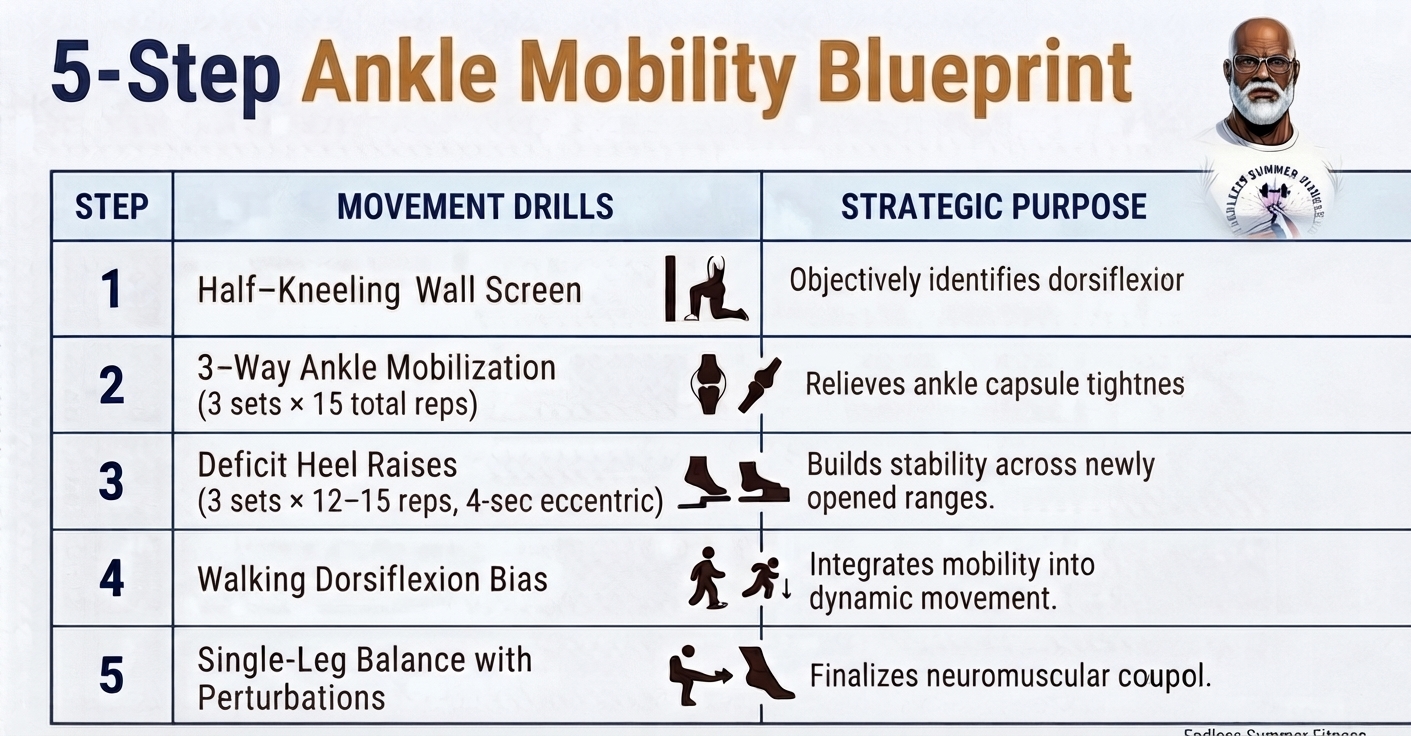
Integrate this movement blueprint into your weekly routine 2 to 3 times per week prior to your walks or workouts:
What to Do Next
Start with STEP 1 today. Screen your ankle dorsiflexion range before your next walk. Identifying your specific restrictions takes under two minutes, giving you a clear, efficient path toward restored movement power and pain-free strides.
Rebuilding your movement confidence is an ongoing process. Unlock your ankles, train with controlled intention, and keep your stride strong for years to come.
Internal Links
References
-
Mitchell, W. K., et al. (2012). Sarcopenia, dynapenia, and the impact of advancing age on human skeletal muscle size and strength; a quantitative review. Frontiers in Physiology, 3, 260. PubMed
-
Zhang, Q., et al. (2024). Effects of peak ankle dorsiflexion angle on lower extremity biomechanics and pelvic motion during walking and jogging. Journal of Biomechanics / PMC, PMC10867967. PMC Article
-
Kim, J. A., et al. (2013). The Effects of Changes of Ankle Strength and Range of Motion on Balance Stability in Elderly. Annals of Rehabilitation Medicine, 37(1), 83–90. PMC Article
-
Vandervoort, A. A., et al. (1992). Age and sex effects on mobility of the human ankle. Applied Physiology, Nutrition, and Metabolism / PubMed, 1730848. PubMed
-
Fong, C. M., et al. (2011). Ankle dorsiflexion range of motion influences dynamic balance and lower extremity kinematics. Journal of Athletic Training, 46(2), 133–140. PMC Article